


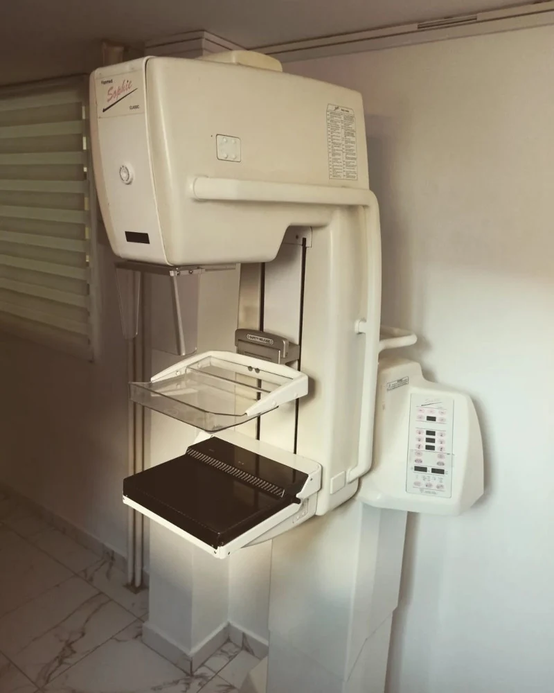
Mammographic examination
The increase in the number of malignant breast diseases has drawn public attention to diagnostic methods in
detection of the disease.
What is mammography?
Mammography is a diagnostic method for detecting breast disease that uses very low doses of X-rays. The examination is performed by our trained radiological technician, and both breasts are imaged from two views. The examination usually takes around ten minutes. The technician must position each breast correctly for imaging, which is not always easy; mutual understanding, cooperation and relaxation are therefore needed from every woman undergoing imaging. Imaging is performed with respect for every individual. As every woman has many questions and uncertainties, discussion and a breast examination before mammography are very important.
Is there an adequate alternative for mammography in early tumour detection?
There is none. Breasts change with age; fatty tissue replaces glandular tissue, and only mammographically do such breasts become better visible for the purpose of distinguishing tumours. The greatest advantage of mammography is the detection of the first signs of hidden disease in the form of microcalcifications. There is a possibility, and it is very often necessary, for our radiologist to supplement the mammographic examination with a targeted ultrasound examination, during which we also use the most modern ultrasound techniques and apparatuses.
Every woman must know that mammography is not performed only once, but the advantage of mammographic imaging lies in monitoring, most often on an annual or biennial basis.
Is the examination painful?
Technicians strive to conduct the examination as quickly, carefully, and painlessly as possible. It can be unpleasant only
compression (pressure) of the breast during imaging, because the breast must be compressed into as thin a section as possible for better imaging. The pressure and imaging last only briefly (around ten seconds). The device we use has so-called softer compression.
Is it necessary and important to have the examination?
Yes, because it is of inestimable importance for the purpose of protecting one's own health.
What harm can radiation exposure cause?
During mammography, a woman receives half the radiation dose of a standard chest X-ray and one twentieth of the dose of a chest CT scan.