



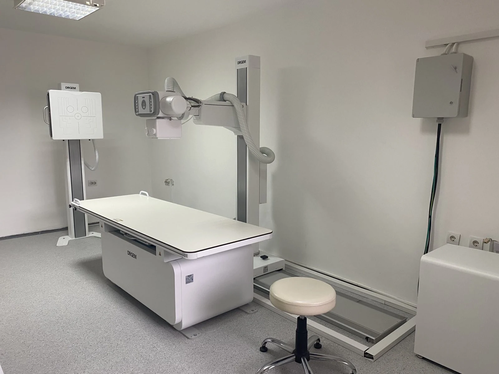
An X-ray examination is a simple and painless method of examination by which images of internal body structures are obtained. It is based on the absorption of ionising X-rays in the tissues of the organism, with the help of an X-ray machine.
Radiography is a painless and non-invasive diagnostic method based on imaging of different parts of the body and organs.
An X-ray machine generates X-rays directed at the part of the body being examined. Denser tissues, such as bones, absorb more X-rays and appear as white areas on the radiograph, while softer tissues, such as organs, appear much darker because they absorb fewer X-rays. The result is an X-ray image. Radiologists analyse the images. Depending on the examination, imaging is performed standing, lying or sitting, and two images are usually taken in two different planes.
Patients are often concerned about the radiation dose they may receive during a diagnostic examination. At our clinic, however, we use a modern X-ray machine that emits minimal radiation.
Radiography is performed as one of the basic examinations when investigating the bone-joint system, so it should be done when there is an injury to some part of the body with suspicion of a bone fracture, joint dislocation, when a deformity or joint swelling is noticed, or when pain appears in different parts of the body (arms, legs, chest, stomach, head).
Radiography is performed by placing the patient, who has prepared for the examination (removed unnecessary clothing and metal objects from the examined part of the body), in front of the plate containing the radiography film. Then the patient is asked to remain motionless during the imaging (e.g., to hold their breath during lung imaging).
Chest X-ray imaging is a fast and painless method by which, with the help of X-rays (x-rays), an image of the chest and the organs located within it – heart, lungs, airways, blood vessels, and lymph nodes – is created. On a chest X-ray image, ribs, sternum, clavicles, and the thoracic part of the spine can also be seen.
Our diagnostic centre possesses the most modern DRGEM GXR series X-ray apparatus, equipped with a high-frequency X-ray generator, which constantly produces high-quality images, thereby ensuring minimal radiation dose during their creation. Our apparatus is designed to provide safety for the technician operating it and comfort for the patient, who deserves it.
We use DICOM archiving in PACS, CD burning and image printing.
Examinations performed on our equipment are:
- X-ray of the lungs
- X-ray of the spine (cervical, thoracic, lumbar)
- X-ray of all joints (knees, hip, ankle, wrist, shoulder, elbow, hand, foot, sacroiliac)
- X-ray of the chest
- X-ray of the abdomen
- X-ray of the urinary tract
- X-ray of the paranasal sinuses
- X-ray of the trachea
- Hysterosalpingography (HSG)